• Skip to Content
  • Skip to Main Navigation
  • Skip to Search

Indiana University Bloomington Indiana University Bloomington IU Bloomington

Open Search
  • About
  • Newsfeed
  • 20 Under 40
  • Photo Essays
  • Alumni Spotlights
  • Short Films
  • Contact Us
  • Give Now

THE COLLEGE

THE COLLEGE THE COLLEGE
  • Home
  • About
  • Newsfeed
  • 20 Under 40
  • Photo Essays
  • Alumni Spotlights
  • Short Films
  • Search
  • Contact Us
  • Give Now
Photo Essays

The Art of Science

Exploring the beauty of the natural world.

By Raymond Fleischmann | Summer 2020

In a 2002 TED Talk, astronaut and physician Mae Jemison spoke about the schism that many perceive between the arts and the sciences. There’s a common belief, she said, that science and the arts are separate, but if you look a little deeper, you’ll find that science and the arts are, in many ways, one and the same.

“Science provides an understanding of a universal experience,” Jemison said in that lecture, “and arts provide a universal understanding of a personal experience.”

When it comes to the work and research of Indiana University faculty and alumni, science and art converge within a range of disciplines, from photography and astronomy to color theory and molecular biology. We recently spoke to four scientists whose work combines the artistic with the scientific, and the result is an inspiring exploration of nature on scales both massive and miniature. See some of their work below.

Every night, College alumnus Mike Weasner (B.S. ’70, Astrophysics) explores the convergence of art and science from his own backyard. After leading a lengthy career in the U.S. Air Force that included time as a fighter pilot, an instructor pilot, and a manager in the Air Force’s space shuttle program, Weasner has turned his full attention to a lifelong passion: astrophotography. He operates a personal observatory from his property near Tucson, Ariz., where he captures images of celestial objects both within our solar system and far beyond it. He’s one of the world’s foremost experts when it comes to the popular Meade ETX Astro Telescope — and for many years operated a related online resource for amateur astronomers — and he played a crucial role in getting Arizona’s Oracle State Park designated as an International Dark Sky Park.

After years of practicing astrophotography, Weasner finds himself returning again and again to certain stars, galaxies, and nebulae. And yet each return to a particular celestial object brings with it the opportunity for new compositions and aesthetic considerations.

“I’ve looked at these objects many times,” Weasner says, “and I’ve often photographed them many times, so when I take another photograph of these celestial objects, I’m trying to get more detail out of the picture or get an object’s position juxtaposed with something else that will create an interesting composition. When I capture sky pictures, for instance — whether it’s the Milky Way or star trails — I’ll often foreground an object that lends more depth to the picture and also grounds the person who’s looking at the photograph. It allows them to say, ‘Hey, this is something real and relatable to our planet Earth,’ whereas if you’re looking at a picture of a galaxy isolated within a telescope, you can’t really relate to that object in the same way.”

Weasner stresses, too, that capturing certain natural phenomena can be an elusive endeavor. Total solar eclipses, for instance, don’t happen every day. The eclipse that occurred in North America in August 2017 was Weasner’s first successfully photographed total solar eclipse, but not his first attempt.

“My first attempt was in March 1970,” he says, “when I was a senior at IU. A bunch of us drove down to Florida to observe the eclipse, but we got totally clouded out and couldn’t see anything.”

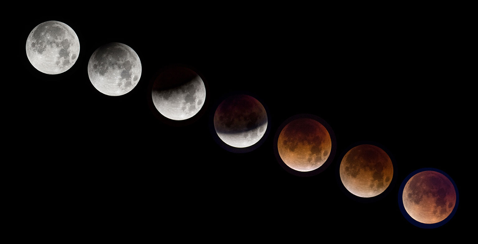
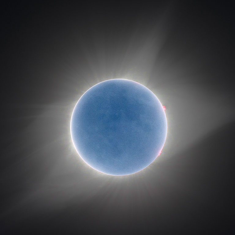
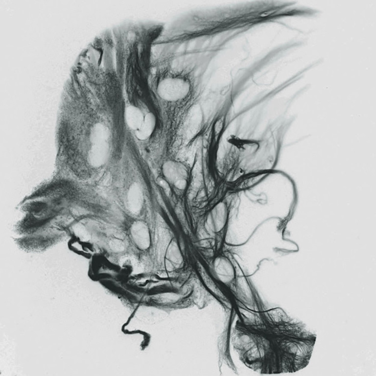
Weasner captured a variety of images from the 2017 event, two of which are pictured above.

“The first image, the one in black-and-white, is a very short exposure, which I used in order to capture the bright streamers within the sun’s corona,” Weasner says. “The second image isn’t filtered or artificially colored or anything like that; rather, it’s an image that shows how the moon is being illuminated by earthshine. During a total solar eclipse, your eye can’t see it, but if you do a long exposure, you can bring out the moon being lit up by the reflection off the Earth’s water and clouds and whatever part of the Earth is facing the moon at that time. In that second image, too, at about two o’clock and four o’clock, you can see several pinkish areas. These are actually huge, massive explosions of gas being ejected off the sun and then falling back into the sun. It’s only during a total eclipse that you can see these prominences without special filtration or other more advanced technology.”

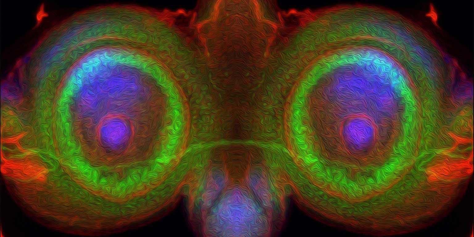
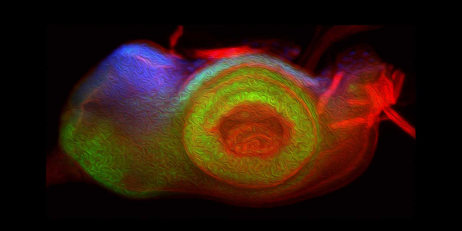
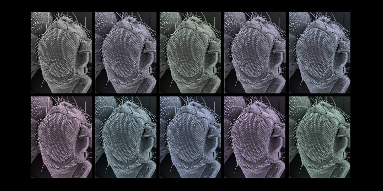
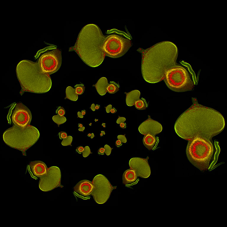
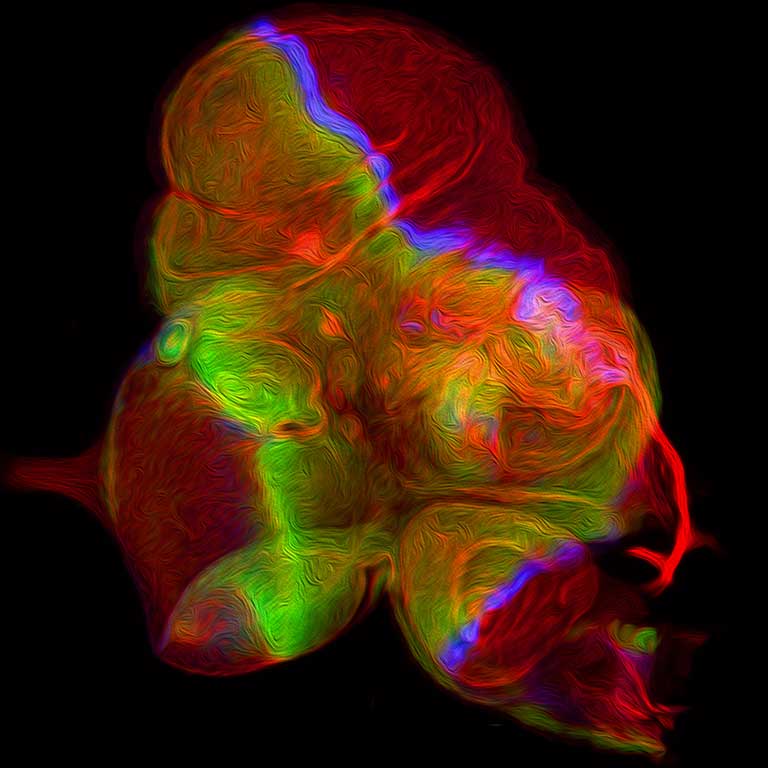
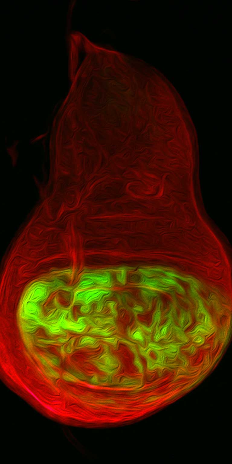
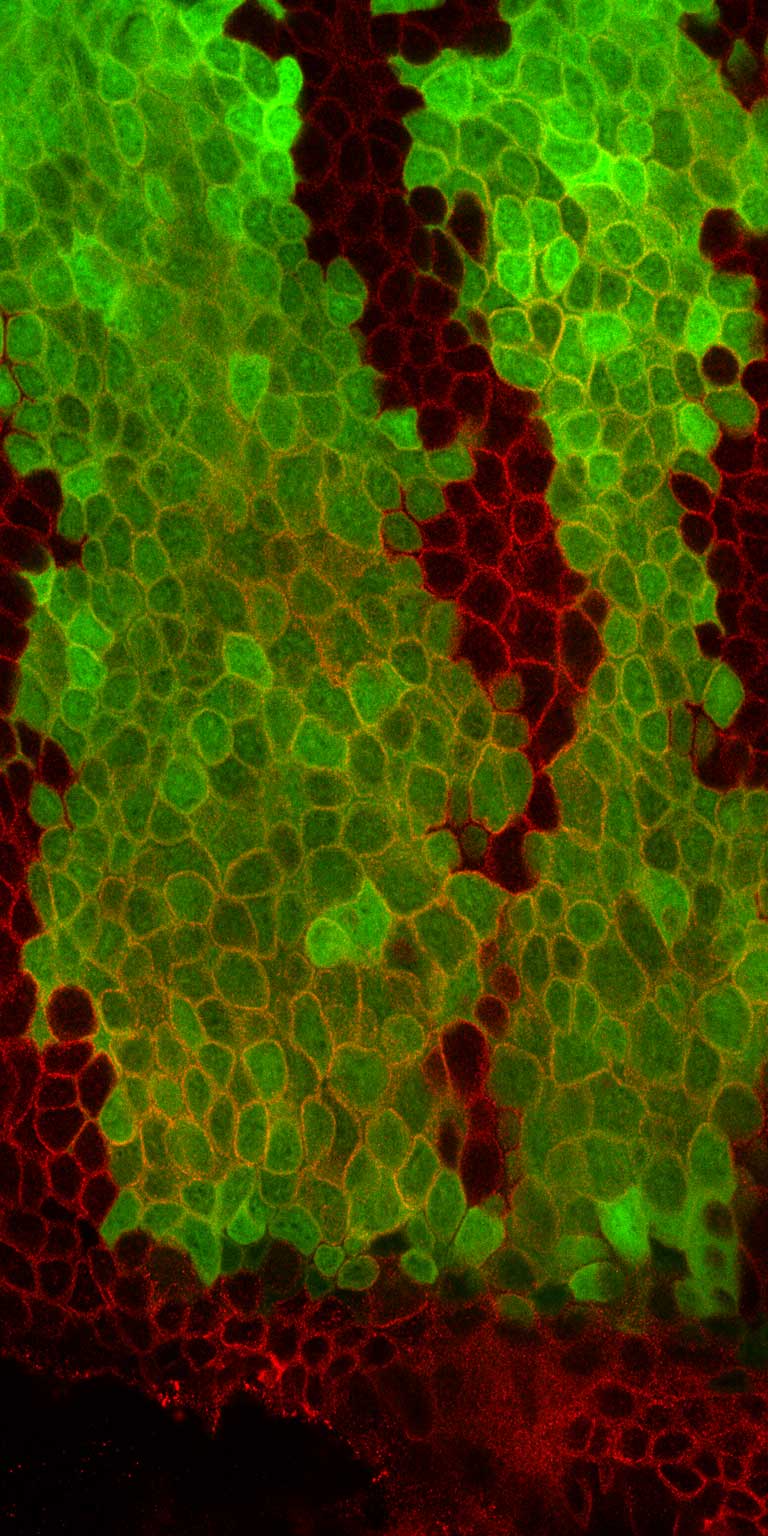
But, of course, the sciences are just as concerned with the miniscule as they are with the massive. Justin Kumar, a developmental biologist and a professor in the College’s Department of Biology, has spent decades studying how the compound eye of the fruit fly, Drosophila melanogaster, acquires its cellular identity and pattern. As an undergraduate studying with molecular biologist Karl Fryxell at the University of California, Riverside, Kumar found a lifelong area of study — and a source of art — at the end of a microscope.

“I become enamored by the simple yet beautiful structure of the Drosophila compound eye,” Kumar recalls. “It contains approximately 750 small unit eyes called ommatidia that are arranged in a precise, repetitive, hexagonal array. Its structure is so stereotyped and free of mistakes that it has been affectionately called a ‘neurocrystalline lattice.’”

Kumar’s research focuses on a collection of Drosophila in which the eye is missing from adult flies, a mutation that has far-reaching implications for both the study of genes and the application of that research.

“All of the genes that we study in Drosophila have functionally equivalent genes in humans,” Kumar explains. “Mutations with the human orthologs are associated with eye disorders such as aniridia, bilateral anophthalmia, congenital cataracts, and photoreceptor degeneration. The loss of the eyes in patients with bilateral anophthalmia mimics the complete loss of the compound eyes that we see in our flies. The conservation of gene function and the similarity of mutant phenotypes allows us to use the fly as an experimental model system to understand how key genes function to specify and pattern the eye in both flies and humans. The collective hope is that we can contribute to potential genetic cures for some of the congenital disorders that plague humans.”

Throughout his years of conducting this research, Kumar has become an expert in microscopic photography, which in turn has appealed to his artistic side.

“Each microscope that we use to visualize developing or adult eyes of the fruit fly is connected to a computer and equipped with a high-resolution digital camera,” Kumar says. “The accompanying software on the computer allows us to control basic parameters such as image orientation, magnification, exposure, brightness, and contrast. Various colors are then applied using Adobe Photoshop. At this point the image is ready for inclusion in research publications, but for inclusion on my laboratory website and on social media as well as on journal covers, I use sets of effect filters to give an image an artistic quality.

“When assigning colors and filters to an image I try out various combinations. Many of them are equally pleasing to the eye, but there is always one that jumps out to me. I try to find the right color and filter combination that will make me stare at it for hours and entice me to go back and look at it again and again.”

For Roger Hangarter, the convergence of art and science is as much a pursuit of education as it is self-expression. A distinguished professor of biology and the Class of 1968 Chancellor’s Professor of Biology, Hangarter is an expert on chloroplast movements in plants, but his interest in photography encompasses a variety of natural subjects.

“With almost every picture I take,” Hangarter says, “in the back of my mind, I’m always thinking, ‘Is there something that somebody could learn about the world through these photographs?’ Capturing images that make you pause and maybe even investigate them a little more are, I think, very valuable.”

In the second and third images below, for instance, Hangarter captures the frenzied movement of protozoa in a pond.

“Those aren’t scientific photographs,” Hangarter says, “since you can’t even tell what kind of organism is pictured — you could guess, but you couldn’t be sure — but in capturing those photographs in the way that I did, you’re still seeing things that most people miss, like the swarming behavior of the different protozoa and the way that they interact with each other. These are things you don’t always notice, because who sits and stares at pond water? It’s a matter of seeing things a little differently.”

Beyond more traditional wildlife photography, Hangarter’s artwork often employs techniques directly related to his scientific research. The photos below, for instance, aren’t doctored or computer-generated; rather, they’re created by manipulating the movement of chloroplasts within the leaves themselves.

“Each cell of a leaf is filled with chloroplasts,” Hangarter explains, “and the chloroplasts will line up around the edges of the cell depending on light intensity. You can actually see the result of this in living leaves by covering up part of a leaf with black paper and letting it sit out in the sun for half an hour.”

Hangarter employs this natural process to use certain leaves, in effect, as photographic media.

“I develop images, which I print on acetate film in black-and-white, and then put those on top of a leaf that’s then exposed to sunlight for a couple of days,” Hangarter says. “Where it’s been exposed to light, the chlorophyll degrades, but where it’s covered by paper, the chlorophyll is retained a little longer. I typically use leaves that are already undergoing senescence in the fall, so you get additional colors in there, too, from other photochemistry.”

Like Kumar’s fascination with the microscopic world of fruit flies, Alex Straiker has found a world of beauty and intrigue in the most miniature of spaces. A senior research scientist in the Department of Psychological and Brain Sciences, Straiker’s research aims to characterize cannabinoid signaling in the brain and the eye.

“Drugs like THC or CBD plug into that system, but those cannabinoid receptors are found all over the body, and in each place they do something different,” Straiker says. “Right now, I’m excited about cannabinoid regulation of tearing. Cannabis smokers report dry eye, but no one has looked at how exactly that works. Dry eye is a major health issue — something like 20 million Americans are affected by it — and there’s no treatment. We’re finding that the body’s cannabinoid signaling system has a major role in regulating tearing, which is very complex and different in males and females.”

Straiker’s microscopic photography frequently depicts single cells or layers of cells, and the results often feel more like abstract paintings or glasswork than organic imagery. Straiker likens the first photo below to a pane of stained glass, though in fact the image depicts a top-down view of the outer layer of a cornea. The white-on-black image, meanwhile, depicts a nerve cell, and the inverted image depicts a glial cell.

The coloration of Straiker’s images is as much a matter of style as function.

“We use fluorescently labeled tags to show us where the cannabinoid receptors are located,” he explains. “Those tags come in a whole spectrum of colors, but we usually choose red and green, partly because the eye is optimized for seeing those colors, especially green, but also because when the two overlap it shows up as yellow. Often we’re trying to figure out whether something new overlaps with something we already know, but because the signals are digitized, we could — and sometimes do — assign whatever color we want.”

In capturing these images, Straiker is well aware of the aesthetic as well as the scientific.

“It’s a bit like landscape photography,” he says, “where the landscape is just very small. I do have a sense that there is a wealth of beauty locked away in these tiny structures, and I feel fortunate to have a window into that world. But a main reason I collect and share these images is to make science more accessible. People often think of science as cold and sterile, but there’s also beauty.”

Related articles

The Music of Movement
Fashion Forward
On the Air


Raymond Fleischmann

Raymond Fleischmann is director of advancement communications for the College of Arts and Sciences and serves as the primary editor for The College magazine. He holds a B.A. in English and the Individualized Major Program from Indiana University, and an M.F.A. in creative writing from Ohio State University. His first novel, How Quickly She Disappears, was published by Penguin Random House in January 2020, and his short fiction has been published in The Iowa Review, Cimarron Review, The Pinch, River Styx, and Los Angeles Review, among many others. Reach him at rfleisch@indiana.edu or through his website raymondfleischmann.com.

THE COLLEGE social media channels

  • Twitter
  • Facebook
  • Instagram
  • YouTube
  • LinkedIn

Indiana University

Accessibility | College Scorecard | Open to All | Privacy Notice | Copyright © 2025 The Trustees of Indiana University